Elements of morphology: Standard terminology for the lips, mouth, and oral region†‡
Note: Individuals are free to copy, distribute and display this work and to make derivative works for noncommercial purposes so long as the work is given proper attribution per the Creative Commons License 3.0. Any derivative works so made should contain the following legend “This is a derivative work of [full cite], made pursuant to Creative Commons License 3.0. It has not been reviewed for accuracy or approved by the copyright owner. The copyright woner disclaims all warranties.”
How to cite this article: Carey JC, Cohen MM Jr., Curry CJR, Devriendt K, Holmes LB, Verloes A. 2009. Elements of morphology: Standard terminology for the lips, mouth, and oral region. Am J Med Genet Part A 149A:77–92.
Abstract
An international group of clinicians and scientists working in the field of dysmorphology has initiated the standardization of terms used to describe human morphology. The goals are to standardize these terms and reach consensus regarding their definitions. In this way, we will increase the utility of descriptions of the human phenotype and facilitate reliable comparisons of findings among patients. Discussions with other workers in dysmorphology and related fields, such as developmental biology and molecular genetics, will become more precise. Here we summarize the anatomy of the oral region and define and illustrate the terms that describe the major characteristics of the lips and mouth. © 2009 Wiley-Liss, Inc.
INTRODUCTION
This paper is part of a series of six articles defining the morphology of regions of the human body [Biesecker et al., 2009; Hall et al., 2009; Hennekam et al., 2009; Hunter et al., 2009; Allanson et al., 2009b]. The series is accompanied by an introductory article describing the general aspects of this study and the principles used in establishing the definitions [Allanson et al., 2009a]. The reader is encouraged to consult the Introduction when using the definitions.
ANATOMY OF THE LIPS, MOUTH, AND ORAL REGION
General
The appearance of the lips varies with facial movement. Smiling and crying can alter dramatically the shape of the upper lip, as do pursing or pouting. Therefore, the lips must be assessed when the subject has a relaxed (neutral) face: the eyes are open, the lips make gentle contact, and the teeth are slightly separated. The neck, jaw, and facial muscles should not be stretched nor contracted, and the face should be positioned using the Frankfurt horizontal (a line joining the orbitale and the porion) [Farkas, 1981]. Here we define the anatomic features important in proposing the Definitions of the paper.
Anatomy
Lips: The structures that surround the oral aperture (Fig. 1). In the central region their superior border corresponds to the inferior margin of the base of the nose. Laterally, their limits follow the alar sulci and the upper and lower lips join at the oral commissures. The inferior limit of the lips in the central region is the mentolabial sulcus. Anatomically, the philtrum and its pillars are a part of the upper lip. The surface of the lip is comprised of four zones: hairy skin, vermilion border, vermilion and oral mucosa. The normal shape of the lips varies with age, and is influenced by ethnicity.

This drawing depicts the major anatomic landmarks of the lips and mouth (see text).
Vermilion: The red part of the lips (Fig. 1). It is covered with a specialized stratified squamous epithelium, which is in continuity with the oral mucosa of the gingivolabial groove. Confusingly, the vermilion itself is also often referred to as “the lips.”
Vermilion border: The rim of paler skin that demarcates the vermilion from the surrounding skin.
Cupid's bow: The contour of the line formed by the vermilion border of the upper lip. In a frontal view, this line resembles an archer's bow, which curves medially and superiorly from the commissures to the paramedian peaks located at the bases of the pillars of the philtrum (crista philtrae) with an inferior convexity lying between those peaks. The philtrum is the vertical groove in the midline of the upper lip bordered by these lateral pillars (ridges) [Hennekam et al., 2009].
Oral mucosa: Stratified squamous non-keratinized epithelium covering of the inner aspect of the oral cavity [Standing, 2005].
Mouth: The oral aperture that opens into the oral cavity proper [Standing, 2005]. The opening is bounded by the upper and lower vermilion. The cavity comprises the alveolar arches with gums and teeth, the hard and soft palate, and the tongue, anchored to the floor of the mouth (Fig. 2). The oral cavity leads into the oropharynx, bounded by the tonsillar pillars. Standards exist for measuring the length and height of the oral aperture [Farkas, 1981].

This drawing depicts the important features of the oral cavity (see text).
Oral commissure: The place where the lateral aspects of the vermilion of the upper and lower lips join. The cheilion is the anthropological landmark located at this site (see Fig. 1).
Labial fissure: Slit-like space between the lips; the oral vestibule.
Oral Cavity: The space bounded superiorly by the hard and soft palates, laterally by the alveolar processes of the maxillary bone, and inferiorly by the tongue (see Fig. 2).
Alveolar ridge: The U-shaped bony crests of the upper and lower jaw in which the teeth are situated.
Hard palate: Bony anterior two-thirds of the roof of the mouth separating the nasal cavity from the oral cavity. The boundary of the hard and soft palates can be determined by palpation.
Soft palate (velum palatinum): Posterior one third of the palate comprised of a fibromuscular fold of soft tissue suspended from the hard palate and separating the nasal and oral cavities.
Uvula: A conical projection of soft tissue extending inferiorly from the posterior edge of the middle of the soft palate (see Fig. 2).
Gingiva (gums): Dense fibrous tissue covered by mucous membrane overlying the alveolar ridge in which the teeth are situated.
Buccal frenulum: A thin fold of soft tissue extending from the gingiva of the mid-anterior alveolar ridge to the inner surface of the medial part of the upper or lower lip (see Fig. 2).
Lingual frenulum: A thin fold of soft tissue extending from the floor of the mouth to the base of the tongue.
Tongue: Muscular organ of deglutition, speech and taste covered with epithelium and bound to the floor of the mouth.
Teeth: Hard dental structures located on the alveolar ridges and situated in the gingiva. In humans, teeth have two stages, the primary (deciduous) and the secondary (permanent, adult).
LIPS: DEFINITIONS
Commissural Pit
Definition: Depression located at an oral commissure (Fig. 3). objective

Commissural pit. These are always located at the same position at the corners of the oral aperture.
Comments: This pit has no relationship to a Lip pit.
Cupid's bow: see Cupid's bow, exaggerated
Cupid's bow, accentuated: see Cupid's bow, exaggerated
Cupid's Bow, Absent
Definition: Lack of paramedian peaks and median notch of the upper lip vermilion (Fig. 4). objective

Absent Cupid's bow. This feature is often associated with a thin vermilion of the upper lip as in this child.
Comment: This bow is often absent in a Thin vermilion of the upper lip, but that should be assessed separately. This finding is commonly associated with Smooth philtrum, but that should be coded separately [Hennekam et al., 2009].
Cupid's Bow, Exaggerated
Definition: More pronounced paramedian peaks and median notch of the Cupid's bow (Fig. 5). subjective

Exaggerated Cupid's bow.
Comment: This may be associated with a Deep philtrum, [Hennekam et al., 2009] but that finding should be coded separately.
Synonym: Cupid's bow, accentuated
Replaces: Cupid's bow (used without adjective)
Lip fistula: see Lip pit
Lip Freckle
Definition: Increased focal pigmentation of the vermilion of the lips (Fig. 6). subjective

Freckling of the vermilion of the lower lip.
Comment: Lip freckles may be accompanied by Perioral hyperpigmentation, but this should be assessed separately. Lentigo is commonly used as a synonym for freckle in reference to the vermilion, but these are distinct terms when referring to the skin.
Synonym: Lip lentigo
Lip, coarse: see, Vermilion, upper lip, thick
Lip, full: see Vermilion, upper lip, thin
Lip, lentigo: see Lip freckle
Lip, lower drooping: see Vermilion, lower lip, everted
Lip, lower full: see Vermilion, lower lip, thick
Lip, lower thick: see Vermilion, lower lip, thick
Lip Pit
Definition: Depression located on the vermilion of the lower lip, usually paramedian (Fig. 7). objective

Typical lip pits. Note: these are usually just lateral to the midline.
Comments: A lip pit may be connected by a fistula to mucous minor salivary glands in the lower lip. In addition, a lip pit may on occasion be seen with a surrounding tissue elevation (mound). Pits located at the labial commissure (cheilion) are distinct from lip pits; see Commissural pit.
Synonym: Lip fistula
Lip, thick: see Vermilion, upper lip, thin Lip, upper thin: see Vermilion, upper lip, thin
Mouth, tented: see Vermilion, upper lip, tented
Nasolabial crease, hypoplastic: see Nasolabial fold, underdeveloped
Nasolabial crease, prominent: see Nasolabial fold, prominent
Nasolabial crease, underdeveloped: see Nasolabial fold, underdeveloped
Nasolabial Fold, Prominent
Definition: Exaggerated bulkiness of the crease or fold of skin running from the lateral margin of the nose, where nasal base meets the skin of the face, to a point just lateral to the corner of the mouth (cheilion, or commissure) (Fig. 8). subjective

Prominent nasolabial fold.
Comments: Increasing prominence with age is usual. See Allanson et al. 2009b.
Synonym: Nasolabial crease, prominent
Nasolabial Fold, Underdeveloped
Definition: Reduced bulkiness of the crease or fold of skin running from the lateral margin of the nose, where nasal base meets the skin of the face, to a point just lateral to the corner of the mouth (cheilion or commissure) (Fig. 9). subjective

Underdeveloped nasolabial fold.
Comments: See Allanson et al. 2009b.
Synonym: Nasolabial crease, underdeveloped
Replaces: Nasolabial crease, hypoplastic; Nasolabial fold, hypoplastic
Perioral Hyperpigmentation
Definition: Increased pigmentation, either focal or generalized, of the skin surrounding the vermilion of the lips (Fig. 10). subjective
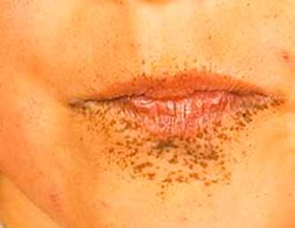
Perioral hyperpigmentation.
Comment: Periorbital hyperpigmentation may be accompanied by Lip freckles, but this should be assessed separately.
Vermilion border, thin: see Vermilion, upper lip, thin
Vermilion, Lower Lip, Everted
Definition: Inner aspect of the lower lip vermilion (normally apposing the teeth) visible in a frontal view (Fig. 11). subjective
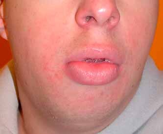
Everted vermilion of the lower lip. The designation pouting lower lip is sometimes used but this is a functional term.
Comments: In frontal view, with the face relaxed, the apparent height of the lower lip vermilion is excessive and the lower incisors may be visible. On profile view, the vermilion is more convex than usual. An everted lower lip may be viewed as “pouting,” but this designation is a functional term.
Replaces: Drooping lower lip
Vermilion, Lower Lip, Thick
Definition: Height of the vermilion of the lower lip in the midline more than 2 SD above the mean (Fig. 12). objective
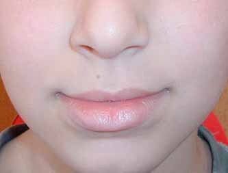
Thick vermilion of the lower lip.
OR apparently increased height of the vermilion of the lower lip in the frontal view. subjective
Comments: Normal values for the height of the vermilion are available [Farkas, 1981] but measurements are not commonly used. Most clinicians determine this feature subjectively. The lower lip is typically thicker than the upper one. The height of the vermilion of the lower lip varies among ethnic groups, and the vermilion should be compared to a population of same ethnic background. When the vermilion is thick, it is more convex and more everted than usual on profile view, but that should be assessed separately.
Replaces: Thick lower lip; Full lower lip
Vermilion, Lower Lip, Thin
Definition: Height of the vermilion of the medial part of the lower lip more than 2 SD below the mean (Fig. 13). objective
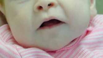
Thin vermilion of the lower lip.
OR apparently reduced height of the vermilion of the lower lip in the frontal view. subjective
Comment: Normal values for the height of the vermilion are available [Farkas, 1981] but measurements are not commonly used. Most clinicians determine this feature subjectively. The height of the vermilion of the lower lip varies considerably among ethnic groups, and the vermilion should be compared to a population of same ethnic background. If the lower lip vermilion is thin, the inferior border of the vermilion is less curved, and on a profile view, the lower lip vermilion is less convex than usual.
Vermilion, Upper Lip, Everted
Definition: Inner aspect of the upper lip vermilion (normally apposing the teeth) visible in a frontal view (Fig. 14). subjective
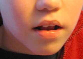
Everted vermilion of the upper lip.
Comments: In frontal view, with the face relaxed, the apparent height of the upper lip vermilion is excessive and the upper incisors may be visible. On profile view, the vermilion is more convex than usual. An everted upper lip may be associated with a short philtrum, and may be secondary to protruded upper teeth, but these should be assessed and described separately.
Vermilion, Upper Lip, Tented
Definition: Triangular appearance of the oral aperture with the apex in the midpoint of the upper vermilion and the lower vermilion forming the base (Fig. 15). subjective
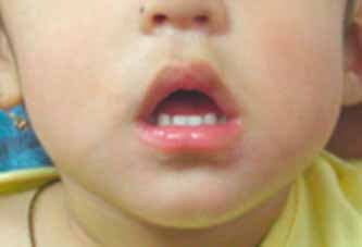
Tented vermilion of the upper lip.
Comment: This finding is distinguished from an Exaggerated Cupid's bow by the alteration of the shape of the oral aperture.
Replaces: Tented mouth
Vermilion, Upper Lip, Thick
Definition: Height of the vermilion of the upper lip in the midline more than 2 SD above the mean (Fig. 16). objective OR
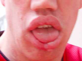
Thick vermilion of the upper lip. This is the preferred designation rather than coarse or full lip since the described feature is the vermilion of the lip, not the lip itself.
apparently increased height of the vermilion of the upper lip in the frontal view. subjective
Comments: Normal values for the height of the vermilion are available [Farkas, 1981], but measurements are not commonly used. Most clinicians determine this feature subjectively or utilize the Likert scale of Astley and Clarren 2000 (Fig. 17). The vermilion of the upper lip varies considerably among ethnic groups, and the vermilion should be compared to a population of same ethnic background. The thickness of the upper lip vermilion is sensitive to the facial expression. On profile view, a thick vermilion is more convex than usual.

This figure displays the commonly used Likert scale, 1–5, that ranges from the Thick vermilion of the upper lip (1) to Thin vermilion; in a Caucasian (A) and an African-American (B) (see text). Note as well the philtral scale from 1 to 5 [see Hennekam et al., 2009].
Replaces: Coarse lip; Thick lip; Full lip
Vermilion, Upper Lip, Thin
Definition: Height of the vermilion of the upper lip in the midline more than 2 SD below the mean (Fig. 18). objective
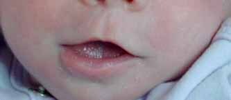
Thin vermilion of the upper lip. Note the absent Cupid's bow as well.
OR apparently reduced height of the vermilion of the upper lip in the frontal view. subjective
Comments: Normal values for the height of the vermilion are available [Farkas, 1981], but measurements are not commonly used. Most clinicians determine this feature subjectively or use the Likert scale for Caucasians and African Americans [Astley and Clarren, 2000] (see Fig. 17). The height of the vermilion of the upper lip varies among ethnic groups, and the vermilion should be compared to a population of same ethnic background. The thinness of the upper lip vermilion is sensitive to facial expression (see Anatomy Section). On profile view, a thin vermilion is less convex than usual. A thin upper lip vermilion may be associated with a smooth philtrum and an absence of the Cupid's bow, but these should be assessed separately.
Replaces: Thin vermilion border; Thin upper lip
MOUTH: DEFINITIONS
Alveolar ridge fusion: see Fibrous syngnathia
Fibrous Syngnathia
Definition: Complete or nearly complete soft tissue fusion of the alveolar ridges (Fig. 19). subjective
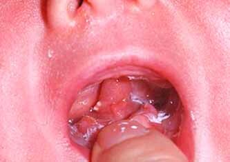
Fibrous syngnathia. See Oral synechiae as well.
Comments: This finding is associated with severely reduced mobility, or lack of mobility, between the upper and lower jaws. This finding is the severe end of a spectrum that includes Oral synechiae.
Synonym: Fusion of the alveolar ridges
Hyperpigmentation, Intra-Oral
Definition: Increased pigmentation, either focal or generalized, of the oral mucosa (Fig. 20). subjective

Intra-oral hyperpigmentation.
Comment: Pigmentation of alveolar ridges is common in people with dark skin pigmentation. This term encompasses a range of pigmentary findings, from freckles to generalized hyperpigmentation.
Macrostomia: see Mouth, wide
Microstomia: see Mouth, narrow
Mouth, carp: see Mouth, downturned corners of
Mouth, Downturned Corners of
Definition: Oral commissures positioned inferior to the midline labial fissure (Fig. 21). subjective
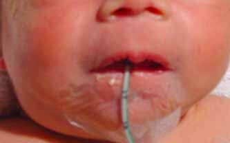
Downturned corners of the mouth. Here the oral commissures are positioned inferior to the midline labial fissure.
Comment: This finding should be assessed with the mouth closed, the lips in relaxed contact, and the face relaxed. The finding may be difficult to assess if the lower lip is enlarged.
Replaces: Carp mouth; Fish mouth (pejorative terms)
Mouth, fish: see Mouth, downturned corners of
Mouth, Narrow
Definition: Distance between the commissures more than 2 SD below the mean (Fig. 22). objective
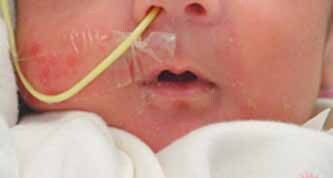
Narrow mouth.
or apparently decreased width of the oral aperture. subjective
Comment: The width of the mouth varies with facial movement and must be assessed when the subject has a relaxed (neutral) face. This term replaces microstomia, small oral aperture, and small mouth because the reduced opening of the mouth is secondary to reduced width.
Replaces: Microstomia; Small oral aperture; Small mouth
Mouth, Upturned Corners of
Definition: Oral commissures positioned superior to the midline labial fissure (Fig. 23). subjective
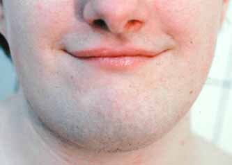
Upturned corners of the mouth.
Comment: This finding should be assessed with the mouth closed, the lips in relaxed contact, and the face relaxed. The finding may be difficult to assess if the upper lip is enlarged.
Mouth, Wide
Definition: Distance between the oral commissures more than 2 SD above the mean. objective OR
apparently increased width of the oral aperture (Fig. 24). subjective

Wide mouth. This width of the oral aperture can be easily measured.
Comment: The width of the mouth varies with facial movement and must be assessed when the subject has a relaxed (neutral) face. This term replaces macrostomia, large mouth, and large oral aperture because these terms imply a wide and open mouth. The term should not be used to describe a patient with a lateral oral cleft.
Replaces: Macrostomia; Large mouth; Large oral aperture
Oral aperture, small: see Mouth, narrow
Oral Frenulum, Accessory
Definition: Extra fold of tissue extending from the alveolar ridge to the inner surface of the upper or lower lip (Fig. 25). objective
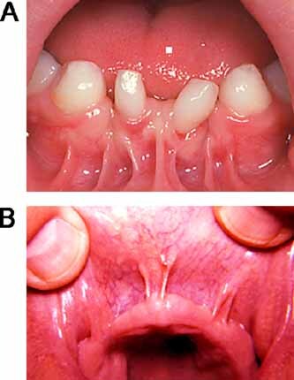
Accessory oral frenulum of the lower (A) and upper lips (B) in different patients.
Comment: This finding is assessed by gently retracting the oral mucosa from the alveolar ridge. Typically there is a single maxillary and a single mandibular frenulum located in the midline between the two central incisors. Abnormalities of the alveolar ridges may accompany accessory frenula, but these should be assessed separately.
Synonyms: Supernumerary oral frenulum; Extra oral frenulum
Oral frenulum, extra: see Oral frenulum, accessory
Oral frenulum, supernumerary: see Oral frenulum, accessory
Oral Synechia
Definition: Fibrous band between the mucosal surfaces of the upper and lower alveolar ridges (Fig. 26). objective
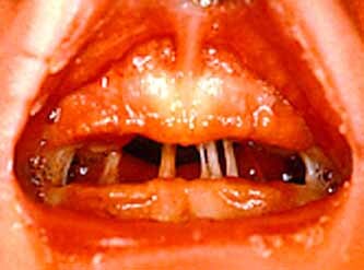
Oral synechiae. Note the obvious fibrous bands.
Comment: These bands must be distinguished from synechiae between the tongue and palate (glossopalatal ankylosis) and from synechiae arising from the floor of the mouth (as in the subglossopalatal membrane), oropharyngeal isthmus (as in persistent buccopharyngeal membrane) or from the lower lip [Gorlin et al., 2001]. If there is a complete soft tissue contiguity between the upper and lower alveolar ridges, the term, Fibrous syngnathia should be used instead.
Vermilion, Upper Lip, U-Shaped
Definition: Gentle upward curve of the upper lip vermilion such that the center is placed well superior to the commissures (Fig. 27). subjective
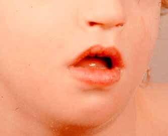
U-shaped vermilion of the upper lip. Note the contour of the vermilion and the shape of the oral aperture.
Comment: The U-shaped upper vermilion is a more rounded version of the Tented upper lip vermilion. In U-shaped upper vermilion there is loss of the central groove of the Cupid's bow.
Replaces: Carp mouth; Fish mouth (pejorative terms); U-shaped mouth
ORAL CAVITY: DEFINITIONS
Aglossia: see Tongue, small
Alveolar ridge hypertrophy: see Alveolar ridge overgrowth
Alveolar Ridge Overgrowth
Definition: Increased width of the alveolar ridges (Fig. 28). subjective
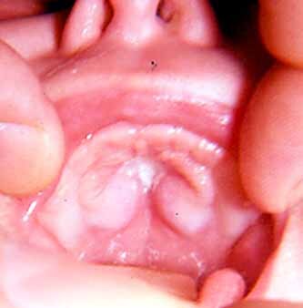
Alveolar ridge overgrowth. Note the increased width of the alveolar ridge.
Comments: This finding may or may not be accompanied by increased height of the alveolar ridge. This is not to be confused with Prominent palatal ridges or Gingival overgrowth. This distinction of gingival from alveolar ridge overgrowth may be difficult, especially in milder degrees of the finding.
Replaces: Alveolar ridge hypertrophy
Ankyloglossia
Definition: Short or anteriorly attached lingual frenulum associated with limited mobility of the tongue (Fig. 29). subjective
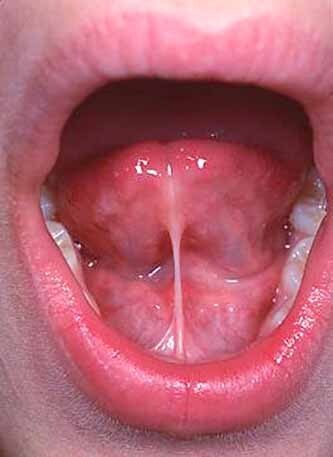
Ankyloglossia. Note the accompanying short lingual frenulum and mild indentation of the tongue tip.
Comment: The anterior third of the tongue is usually free or is partially attached to the floor of the mouth by the lingual frenulum. There is a spectrum ranging from fusion of the tongue to the floor of the mouth (“ankyloglossia inferiorum”) to a lingual frenulum that is short or anchored toward the tip of the tongue (“tongue tie”). Ankyloglossia may be associated with a mild indentation of the tip of the tongue, which should not be coded as a Bifid tongue.
Replaces: Ankyloglossia inferiorum; tongue tie
Anodontia: see Oligodontia
Central Incisor, Single Maxillary
Definition: Presence of one maxillary central incisor positioned in the midline (Fig. 30). objective
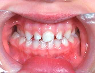
A single central maxillary incisor. The lateral incisors are normal making the recognition of this feature difficult on first glance.
Comment: If a single maxillary central incisor is present but not positioned in the midline, this could be hypodontia (see Ologodontia), but this cannot be evaluated without a radiograph.
Dental Crowding
Definition: Overlapping teeth within an alveolar ridge (Fig. 31). subjective
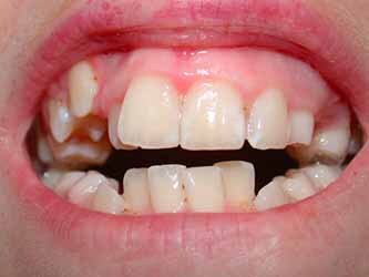
Dental crowding.
Comment: This is a bundled term. There is a discrepancy in the size or number of the teeth compared to the size of the alveolar ridges.
Diastema
Definition: Increased space between two adjacent teeth in the same dental arch (Fig. 32). subjective
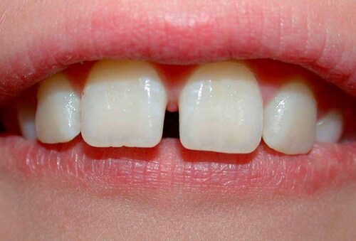
Diastema. See widely spaced teeth as well.
Comments: Usually there is contact between the lateral aspects of the permanent teeth, at their broadest point. Diastema can apply to any pair of teeth and the term should be modified by a descriptor of the involved teeth. This descriptor must be distinguished from Widely spaced teeth.
Diastemata, multiple: see Teeth, widely spaced
Double tooth: see Teeth, fused
Eruption, Advanced
Definition: Tooth eruption more than 2 SD earlier than the mean eruption age. objective
Comment: There are established norms for the timing of eruption in both deciduous and permanent teeth [Garn and Rohmann, 1966; Lunt and Law, 1974; McDonald et al., 2004]. Eruption is defined by the appearance of a tooth that has pierced the gum.
Eruption, Delayed
Definition: Tooth eruption more than 2 SD beyond the mean eruption age. objective
Comment: This term should not be used in a patient with Gingival overgrowth. There are established norms for the timing of eruption in both deciduous and permanent teeth [Garn and Rohmann, 1966; Lunt and Law, 1974; McDonald et al., 2004]. Eruption is defined by the appearance of a tooth that has pierced the gum.
Gingival hyperplasia: see Gingival overgrowth
Gingival hypertrophy: see Gingival overgrowth
Gingival Overgrowth
Definition: Thickening of the soft tissue overlying the alveolar ridge (Fig. 33). subjective
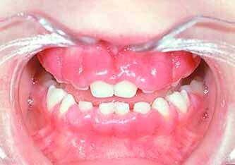
Gingival overgrowth. Note the difference between this finding and overgrowth of the alveolar ridge.
Comments: The degree of thickening ranges from involvement of the interdental papillae alone to gingival overgrowth covering the entire tooth crown.
Replaces: Gingival hypertrophy, Gingival hyperplasia
Glossoptosis
Definition: Posterior displacement of the tongue into the pharynx (Fig. 34). subjective
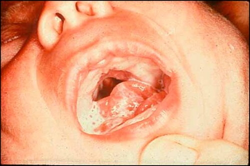
Glossoptosis. Note the tongue's posterior placement in the oral cavity and the presence of the formula. (Figure courtesy of Bryan Hall.)
Comment: Presumably, use of the suffix “ptosis” refers to the situation where the patient is supine, and the displacement is downward. Strictly speaking, the term glossoptosis indicates “falling” of the tongue and thus can also be forward displacement; however by convention it is only used for backward displacement. Glossoptosis may cause obstruction of the airway.
Hypodontia: see Oligodontia
Hypoglossia: see Tongue, small
Macrodontia
Definition: Mesiodistal tooth diameter (width) more than 2 SD above mean for age (Fig. 35). objective OR
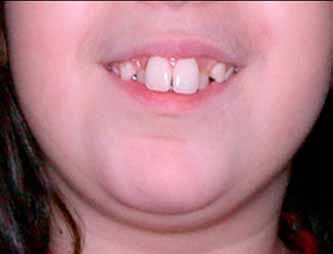
Macrodontia. The tooth width is easily measured.
apparently increased maximum width of the tooth. subjective
Comment: The standard reference has means and standard deviations by gender [Moyers et al., 1976].
Macroglossia: see Tongue, large
Microdontia
Definition: Mesiodistal tooth diameter (width) more than 2 SD below mean (Fig. 36). objective
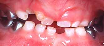
Microdontia.
OR apparently decreased maximum width of tooth. subjective
Comment: Standard reference has means and standard deviations by gender [Moyers et al., 1976]. In microdontia, the gaps between the teeth, particularly the anterior upper and lower teeth, are increased, creating Diastemata. This should be assessed and coded separately.
Microglossia: see Tongue, small
Oligodontia
Comment: The term is not defined here since the finding requires a radiograph, as is true for anodontia and for the other designation of tooth agenesis, hypodontia. The terms hypodontia and oligodontia are sometimes used interchangeably in the literature while on other occasions hypodontia is used for selective agenesis of six or less missing teeth while oligodontia is applied when there are more than six missing teeth. Tooth agenesis or oligodontia/hypodontia can be mistaken for delayed eruption and again a radiograph is needed for diagnosis. Absence of teeth may be congenital (tooth agenesis) or acquired. The incidence of congenital absence of teeth is different depending on the type and position of the tooth [Gorlin et al., 2001].
Open Bite
Definition: Visible space between the dental arches in occlusion (Fig. 37). objective
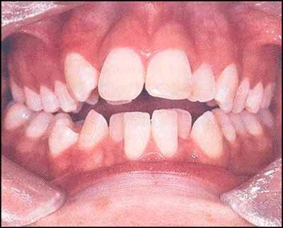
Open bite. Note the space between the dental arches. (Figure courtesy of Duane Yamashiro.)
Comments: An open bite produces an absence of vertical overlap of the two dental arches. It may be associated with malocclusion, but this should be coded separately. Open bite can be accompanied by malocclusion, which is a complex bundled term. The Angle classification of malocclusion (Classes I–III) is widely used in the orthodontics community [Moyers, 1973] for the characterization of malocclusion.
Palate, Hard, Short
Definition: Distance between the labial point of the incisive papilla to the midline junction of the hard and soft palate more than 2 SD below the mean (Fig. 38). objective
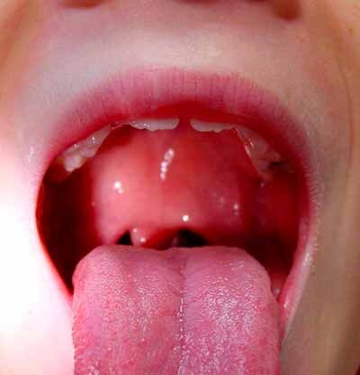
Short hard palate. (Figure courtesy of Alan Rope.)
OR apparently decreased length of the hard palate. subjective
Comment: Objective measurement of the hard palate requires special instrumentation [Hall et al., 2006]. A short hard palate may be associated with velopharyngeal incompetence.
Replaces: Short palate; hypoplastic palate
Palate, short: see Palate, hard, short
Palate, High
Definition: Height of the palate more than 2 SD above the mean. objective OR
Palatal height at the level of the first permanent molar more than twice the height of the teeth (Fig. 39). subjective
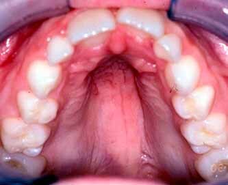
High palate. Note a Narrow palate is a different feature and can produce the false appearance of a high palate.
Comments: The measuring device for this assessment is described in Hall et al. 2006. A high palate is often associated with a narrow palate. However, a narrow palate can easily give a false appearance of a high palate. Height and width of the palate should be assessed and coded separately. We do not recommend the subjective determination because this term can be overused and applied inaccurately.
Synonym: High, arched palate
Palate, high arched: see Palate, high
Palate, hypoplastic: see Palate, hard, short
Palate, Narrow
Definition: Width of the palate more than 2 SD below the mean. objective
OR apparently decreased palatal width (Fig. 40). subjective
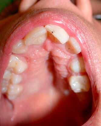
Narrow palate. Note Prominent palatine ridges as well.
Comments: Palatal width is measured as the distance between the maxillary first permanent molar on the right and left sides, at the lingual cervical line, using a specific device. Palate width is typically assessed subjectively in routine clinical practice. Narrowing is often associated with a High palate, but this should be assessed and coded separately. Gingival overgrowth can give the impression of a narrow palate but should be distinguished and coded separately. The term “gothic palate” is used to indicate that the roof of the palate is not round but rather has an inverted V-shape, and therefore, only the upper part of the palate is narrow.
Palate, Submucous Cleft
Definition: Soft palatal defect with intact overlying mucosa comprising two of the following three findings: (1) notching of the posterior border of the hard palate, (2) bifid uvula, or (3) muscular diastasis leading to a midline translucent zone or furrow in the soft palate (Fig. 41). objective
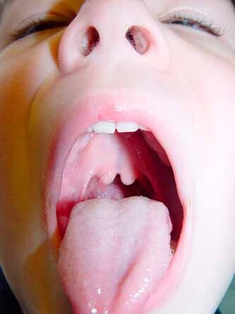
Submucous cleft palate. Note the Cleft uvula and the blue indentation of the soft palate. (Figure courtesy of Robert Shprintzen.)
Comment: The notch of the posterior hard palate can sometimes be palpated [Aase, 1990]. Submucous cleft palate is a bundled term but because of its common usage is included here.
Palatine Ridges, Prominent
Definition: Increased size and/or number of soft tissue folds on the palatal side of the maxillary alveolar ridge (Fig. 42). subjective
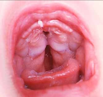
Prominent palatine ridges. Note the more obvious soft tissue ridges and folds.
Comments: Soft tissue folds are typically present on the lateral sides of the palate, especially anteriorly.
Synonym: Prominent lateral palatal ridges; Prominent palatine folds
Teeth, Fused
Comments: Dental fusion or double tooth (the joining of teeth with separate roots) can only be distinguished from gemination (bifid crown, where only one pulp chamber or root canal is present), with radiographic evidence [Cameron and Widmer, 2003]. Therefore this term is not defined here.
Teeth, Widely Spaced
Definition: Increased spaces (diastemata) between most of the teeth in the same dental arch (Fig. 43). subjective
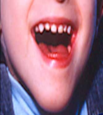
Widely spaced teeth.
Comments: Wide spacing can be secondary to increased room by an unusually large dental arch, microdontia or mixed primary and secondary dentition. It should be carefully noted that slight spacing between the primary teeth is normal, so experience in evaluation is important in determining this feature. This descriptor must be distinguished from Diastema.
Synonym: Multiple diastemata
Tongue, Bifid
Definition: Tongue with a median apical indentation or fork (Fig. 44). objective
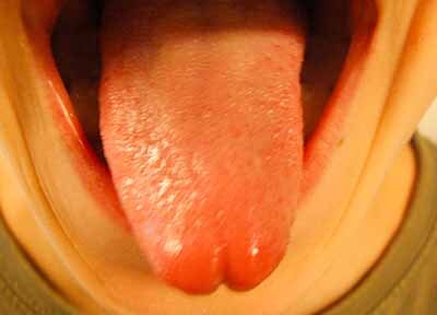
Bifid tongue.
Comments: Bifid tongue can be associated with Ankyloglossia, but this should be assessed and coded separately. Small indentations of the tip of the tongue should not be coded as a bifid tongue.
Tongue, Furrowed
Definition: Accentuation of the grooves on the dorsal surface of the tongue (Fig. 45). subjective
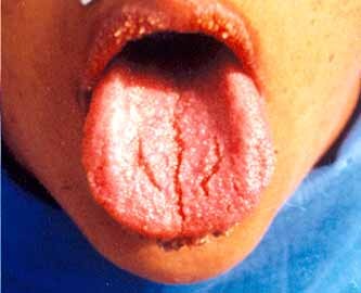
Furrowed tongue.
Comments: Usually there is a midline groove with smaller radiating grooves. The deep furrows may extend to the lateral borders. They may follow a regular geometric pattern or be irregular. A furrowed tongue occurs in 10–25% of individuals but is rare in children.
Synonyms: Prominent tongue grooves
Replaces: Scrotal tongue (pejorative)
Tongue grooves, prominent see Tongue furrowed
Tongue, hyperplasia: see Tongue, large
Tongue, hypertrophy: see Tongue, large
Tongue, hypoplastic: see Tongue, small
Tongue, Large
Definition: Increased length and width of the tongue (Fig. 46). subjective
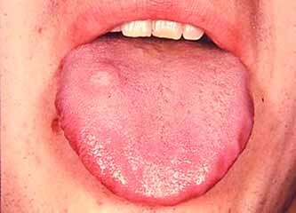
Large tongue.
Comments: Normal standards do not exist. Large size usually leads to protrusion of the tongue. This is an acknowledged bundled term, but due to its frequent usage and relative paucity of situations that would call for separate individual assessments of tongue dimensions, the bundled term is retained. Micrognathia may give the false appearance of a large tongue.
Synonyms: Macroglossia; hyperplasia of the tongue; hypertrophy of the tongue
Tongue, Lobulated
Definition: Multiple indentations and/or elevations on the edge and/or surface of the tongue producing an irregular surface contour (Fig. 47). subjective
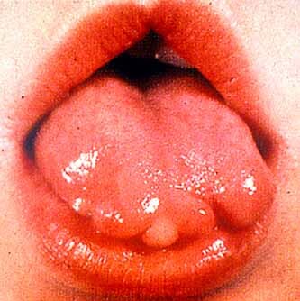
Lobulated tongue.
Comment: Lobulated tongue can bilobed, trilobed, or show multiple lobes.
Tongue, Protruding
Definition: Tongue extending beyond the alveolar ridges or teeth at rest (Fig. 48). subjective
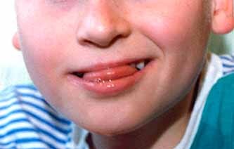
Protruding tongue. Note the position of the tongue.
Comments: Protruding tongue may or may not be associated with a Large tongue, and that finding should be assessed and coded separately.
Tongue, rudimentary: see Tongue, small
Tongue, scrotal: see Tongue, furrowed
Tongue, Small
Definition: Decreased length and width of the tongue (Fig. 49). subjective
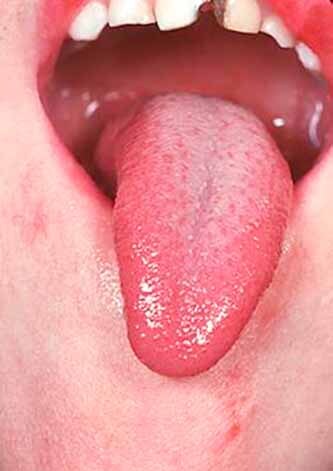
Small tongue.
Comment: Normal standards do not exist. The term “aglossia” is often used for extremely small tongue, but a nubbin of tongue tissue is almost always present and aglossia in sensu strictu is extremely rare. This is an acknowledged bundled term, but due to its frequent usage and relative paucity of situations that would call for separate individual assessments of tongue dimensions, the bundled term is retained.
Synonyms: Microglossia; hypoglossia; rudimentary tongue
Replaces term: Aglossia; hypoplastic tongue; hypotrophic tongue
Tongue, Smooth
Definition: Glossy appearance of the entire tongue surface (Fig. 50). subjective

Smooth tongue. Note the surface of the tongue in this patient.
Comment: This is due to reduction in number and/or size of the filiform papillae. A geographic tongue has localized areas of smoothening, but not sufficient to warrant use of the term Smooth tongue.
Tooth agenesis: see Oligodontia
Tooth, Natal
Definition: Erupted tooth or teeth at birth (Fig. 51). objective
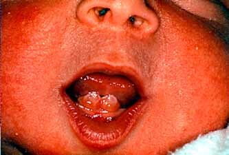
Natal tooth. Note the eruption of this tooth in this newborn infant.
Comment: This is not to be confused with apical dental cysts. Natal teeth most often involve lower central incisors, less often upper central incisors, and rarely first primary molars. Natal teeth occur about once in 3,000 births and are particularly common among some native (First Nation) groups of North America [Mok and Suina, 1986].
Tooth, Premature Loss
Definition: Exfoliation of a primary tooth or teeth earlier than the normal range. objective
Comments: See ranges in Kleigman et al. 2007 and Gorlin et al. 2001. The range of ages in years for normal exfoliation of primary teeth usually precedes the mean age of eruption of each tooth by a year or less.
Tooth, Supernumerary
Definition: Extra tooth or teeth (Fig. 52). objective
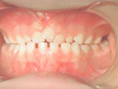
Supernumerary tooth. Note the extra central incisor between the other incisors. Also there are some widely spaced teeth in the lower arch. (Figure courtesy of Duane Yamashiro.)
Comment: The most frequent supernumerary tooth is a mesiodens, which occurs between the two maxillary central incisors. Often it fails to erupt, but creates a large anterior diastema, and would not be detected on physical examination (requires X-ray evaluation). This designation excludes coexistence of primary and permanent dentition due to delayed loss of the former.
Uvula, Absent
Definition: Lack of the uvula (Fig. 53). objective
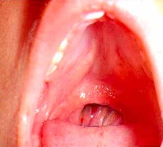
Absent uvula. (Figure courtesy of Robert Shprintzen.)
Comment: Sometimes accompanies a Submucous cleft palate, but this should be coded separately.
Uvula, Broad
Definition: Increased width of the uvula (Fig. 54). subjective

Broad uvula. (Figure courtesy of Robert Shprintzen.)
Comments: This finding often accompanies a Submucous cleft palate, but this should be coded separately. A longitudinal groove indicating incomplete fusion of the two parts of the uvula may be present with a broadened uvula and has sometimes been called abortive cleft uvula.
Uvula, Cleft
Definition: Uvula separated into two parts most easily seen at the tip (Fig. 55). objective
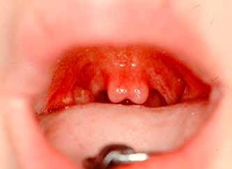
Cleft uvula. Often called a bifid uvula, this feature is a component of a submucous palate but is common as an isolated feature.
Comments: Submucous cleft palate is a distinct entity.
Synonym: Bifid uvula
Uvula, bifid: see Uvula, cleft
Uvula, Long
Definition: Increased length of the uvula (Fig. 56). subjective
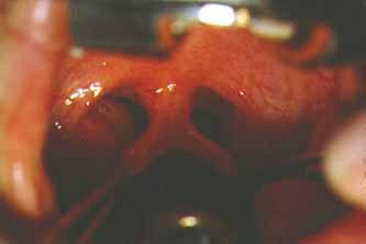
Long uvula. Note the uvula is also cleft. (Figure courtesy of Robert Shprintzen.)
Comments: In clinical practice, the size of the uvula cannot be easily measured and is not static, since it depends on the position of the soft palate, the base of the tongue, and the head. Therefore, judgment of change in length of the uvula depends heavily on the experience of the observer.
Uvula, Narrow
Definition: Decreased width of the uvula (Fig. 57). subjective
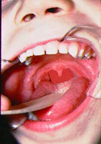
Narrow uvula. (Figure courtesy of Robert Shprintzen.)
Uvula, Short
Definition: Decreased length of the uvula (Fig. 58). subjective

Short uvula. (Figure courtesy of Robert Shprintzen.)
Comments: Objective measurement of the length of the uvula can be determined on a lateral cephalograms. However, in this series we are not relying on radiographs for assessment of findings. In clinical practice, the size of the uvula cannot be easily measured and is not static, since it depends on the position of the soft palate, the base of the tongue, and the head. Therefore, judgment of change in length of the uvula depends heavily on the experience of the observer.
Replaces term: Hypoplastic uvula
Uvula, hypoplastic: see Uvula, short


